
































































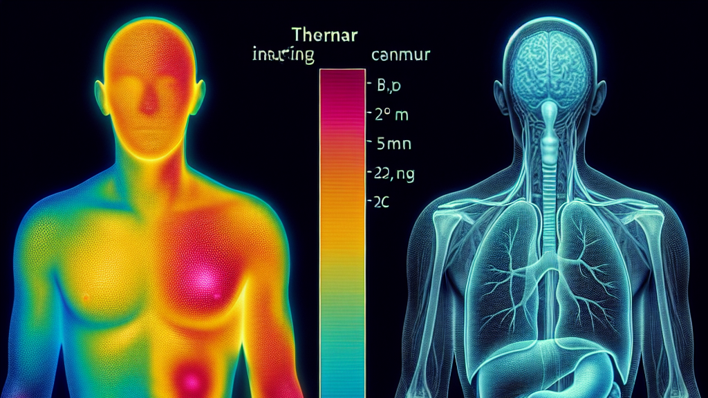
Imagine using heat to reveal hidden secrets of the body. That’s what researchers are doing with a new method to detect tumors. By analyzing temperature patterns on the skin, they can find the size and location of tumors hiding inside. This means easier, non-invasive detection, potentially without the need for complicated procedures like MRIs or X-rays.
The research introduces a novel approach that combines mathematics and thermography, a technique that uses infrared cameras to see temperature variations on the skin. By optimizing a mathematical formula, scientists can estimate where a tumor might be based on these heat variations. This is crucial because the temperature changes could indicate abnormal growths that emit more heat than regular tissue.
Imagine going for a quick scan that feels more like having your picture taken than a medical test. If this method becomes widespread, it could make regular check-ups quicker and more comfortable, making early detection a reality for everyone. This heat-mapping technique might just be the superhero we need in the medical world, protecting us from dangers lurking within before they get a chance to strike.
Infrared thermography, originally developed for military purposes, is now being used to detect tumors by capturing temperature variations across the skin.
FAQs
How does the study propose detecting tumors with heat?
The study introduces a method that uses shape optimization and complex boundary methods to analyze temperature profiles from infrared thermography, focusing on changes that might indicate a tumor’s presence.
Why is infrared thermography used in tumor detection?
Infrared thermography captures temperature variations on the skin, where abnormal heat patterns can indicate underlying issues such as tumors, providing a non-invasive detection method.
What makes this tumor detection method different from traditional imaging techniques?
This method is non-invasive and relies on analyzing thermal images to estimate tumor location and size, potentially reducing the need for MRIs or X-rays.
What is the advantage of using temperature profiles over other methods?
Temperature profiles can quickly indicate areas of concern without exposure to radiation, making them safer and more comfortable for routine check-ups.
Can this method completely replace existing imaging techniques?
While promising, this method isn’t likely to entirely replace existing techniques but could complement them, providing a more comprehensive approach to tumor detection.
Background
In medical diagnostics, identifying a tumor’s size and location is crucial for treatment. Traditional methods like MRIs or X-rays can be invasive or expose patients to radiation. The new method introduced here uses infrared thermography—a way to use cameras to see heat patterns on the skin. By solving complex mathematical puzzles, scientists can determine where a tumor might be hiding based on these patterns.
History
Infrared thermography was first used for military and industrial applications before its potential for medical diagnostics was recognized. Initially utilized to detect skin temperature changes, researchers have developed methods to use this technology for more complex tasks, such as identifying tumors. Over time, advancements in mathematical modeling and computer algorithms allowed for more accurate interpretations of thermal images, leading to innovative diagnostic methods.
Based on “Localization of tumor through a non-conventional numerical shape optimization technique” by Julius Fergy Tiongson Rabago, available on arXiv (arxiv.org/abs/2502.20656), used under CC BY 4.0 (creativecommons.org/licenses/by/4.0/).
