


























































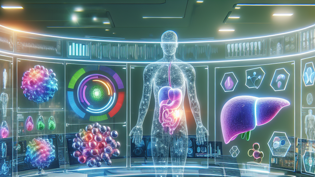
Imagine a future where doctors can quickly and accurately analyze your body fat composition with just a simple scan. This isn’t science fiction—it’s becoming reality thanks to a new A.I. model called Attention GhostUNet++. This technology promises to change the way we look at health risks associated with fat, like diabetes and heart disease.
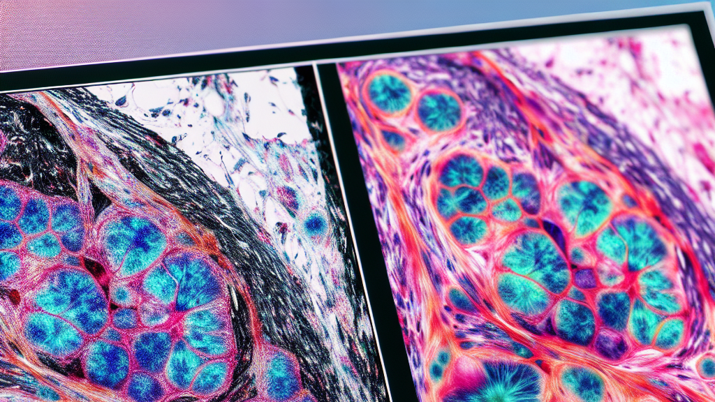
The Attention GhostUNet++ is a cutting-edge deep learning model that has been specifically designed to accurately identify and segment different types of fat in the body and the liver. By focusing on different types of attention mechanisms, this model can pick up on the nuances of where fat is stored, which can be critical in assessing health risks. When evaluated on real-world data, it outperformed older models, showing incredible accuracy.
In the near future, this could mean that your routine health checks include a quick scan that gives you and your doctor detailed information about your visceral and subcutaneous fat levels. The implications are huge, as early detection and precise analysis could lead to better management of diseases like type 2 diabetes and prevent long-term health issues. This technology might soon be a part of regular health check-ups, offering a much clearer picture of your health.
Did you know that precise fat segmentation could help doctors predict diabetes risk more accurately than some traditional tests?
FAQs
What is abdominal adipose tissue segmentation?
Abdominal adipose tissue segmentation is the process of using imaging techniques to accurately separate and identify different types of fat within the abdominal area, like subcutaneous and visceral fat, for better health analysis.
How does the Attention GhostUNet++ model improve health assessments?
The Attention GhostUNet++ model enhances health assessments by using advanced attention mechanisms to accurately segment body fat. This helps in precisely understanding body composition, which is crucial for predicting and managing health risks like cardiovascular disease.
Why is accurate liver segmentation important?
Accurate liver segmentation is important because it helps in understanding liver health and function, which can be indicative of various health conditions. The new model’s precision can assist in better diagnosis and monitoring.
How could this research affect regular health check-ups?
This research could lead to routine health check-ups that include scans providing detailed information on body fat distribution, enabling early detection and better management of potential health risks such as obesity-related diseases.
What are the potential limitations of the new AI model?
While the model excels in accuracy and efficiency, it has minor limitations in boundary detail segmentation, which the developers aim to improve upon in future iterations.
Background
In the realm of health sciences, understanding body fat distribution is crucial because it helps medically assess risks for conditions like type 2 diabetes and cardiovascular disease. Traditional methods of measuring body fat can be invasive or inaccurate, so advanced imaging techniques, like the ones used in this research, have become vital. AI models like the Attention GhostUNet++ use deep learning—a kind of artificial intelligence—to process and interpret complex imaging data accurately. The model uses ‘attention mechanisms’ to focus on important parts of the image, making it highly efficient at recognizing different types of fat and liver tissue.
History
The field of medical imaging has evolved dramatically with advancements in artificial intelligence. Initially, segmentation of body parts in medical images required significant manual effort and expertise. With the introduction of deep learning models like UNet, automatic segmentation became more feasible. The ongoing iterations, like Ghost UNet++ and its latest version, Attention GhostUNet++, build on these foundations by integrating attention mechanisms into the model, which significantly enhances segmentation accuracy and efficiency.
Based on “Attention GhostUNet++: Enhanced Segmentation of Adipose Tissue and Liver in CT Images” by Mansoor Hayat, Supavadee Aramvith, Subrata Bhattacharjee, Nouman Ahmad, available on arXiv (arxiv.org/abs/2504.11491), used under CC BY 4.0 (creativecommons.org/licenses/by/4.0/).