


















Imagine a world where doctors can diagnose cancer almost instantly, catching it in its early stages with incredible accuracy. AI is making this a reality by analyzing real-time images and giving oncologists the power they need to make faster, more precise decisions. This could be a game-changer in the fight against cancer, offering hope to millions of patients worldwide.
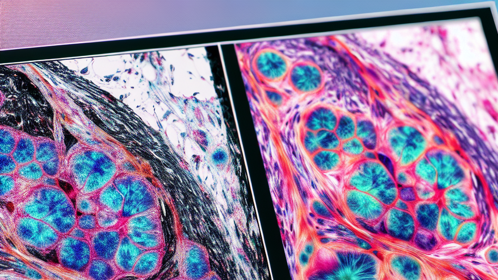
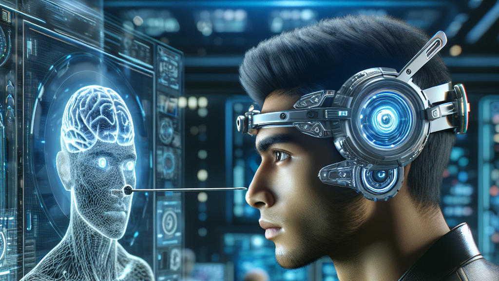
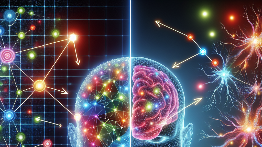
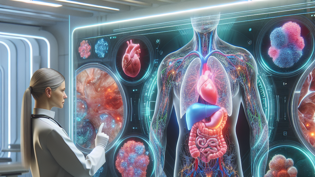
In this groundbreaking research, scientists are exploring how AI can be used to analyze images in real-time to detect various types of cancer. By merging multiple images and using techniques like ultrasound fusion and fluorescence imaging, AI can provide a clearer picture of what’s happening inside the body. It even incorporates electromagnetic needle tracking and neuromorphic architectures to pinpoint cancerous cells, making diagnosis swifter and more reliable.
This technology could transform routine check-ups. Imagine going for a regular mammogram or skin scan and, within minutes, getting a comprehensive analysis that highlights any areas of concern. Not only would this speed up the diagnosis process, but it could also mean starting treatment earlier, potentially saving more lives. The future of cancer detection looks bright, and AI is leading the way.
Did you know? AI can improve cancer diagnosis by acting on image data within seconds, offering potential life-saving insights faster than ever before.
FAQs
How can AI improve cancer diagnosis with real-time imaging?
AI enhances cancer diagnosis by analyzing images instantly, allowing for early detection and precise decision-making. This can improve patient outcomes by enabling faster and more accurate diagnoses.
What techniques are used in AI-based cancer imaging?
AI uses a variety of techniques including ultrasound fusion, fluorescence imaging, and electromagnetic needle tracking to provide detailed, real-time analyses of potential cancerous areas.
Why is early cancer detection important?
Early detection of cancer significantly increases the chances of successful treatment. AI’s ability to quickly and accurately identify cancerous cells enables doctors to start treatment sooner, improving survival rates.
What is image fusion in cancer diagnosis?
Image fusion in cancer diagnosis involves combining different types of imaging data to form a complete and detailed view, helping doctors better identify and understand cancerous tissues.
How does AI compare to traditional cancer diagnosis methods?
AI offers quicker and often more accurate results compared to traditional methods, providing valuable insights during routine check-ups or early symptoms, leading to potentially life-saving early interventions.
Background
AI, or Artificial Intelligence, refers to the capability of a machine to replicate intelligent human behavior. In healthcare, AI can analyze vast amounts of medical imaging data quickly and with high accuracy. Real-time imaging allows doctors to see pictures of the inside of the body as they happen, providing immediate insights. Combining these technologies, doctors can get a full picture of a patient’s health, enabling them to make faster and more informed decisions.
History
Medical imaging began with simple X-rays in the late 19th century. Over the decades, technologies like MRI and CT scans have evolved, becoming critical tools in diagnosing conditions like cancer. More recently, the combination of AI and these imaging techniques has opened new avenues, offering unprecedented speed and accuracy. This research is the next step in this evolution, potentially setting a new standard in cancer diagnostics.
Based on “Advancements in Real-Time Oncology Diagnosis: Harnessing AI and Image Fusion Techniques” by Leila Bagheriye, Johan Kwisthout, available on arXiv (arxiv.org/abs/2503.11332), used under CC BY 4.0 (creativecommons.org/licenses/by/4.0/).