

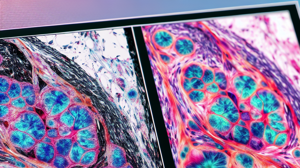






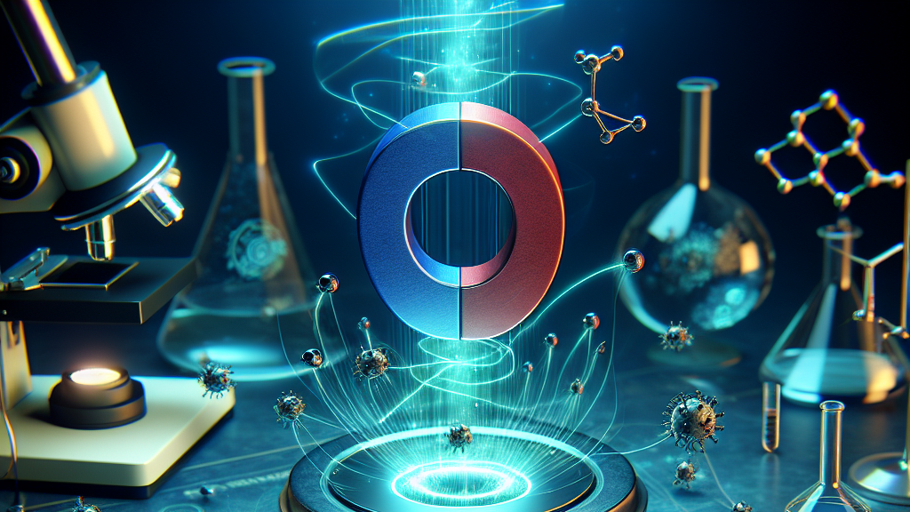

















Imagine being able to see the complex inner workings of a cell in vivid color, much like admiring a beautifully lit city at night. Traditionally, when scientists want to look at cells under a microscope, they use fluorescent labels to make certain parts of the cell light up. But here’s the catch: the number of labels they can use is limited, which means they can only look at a few things at once.
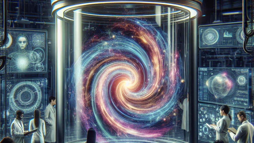
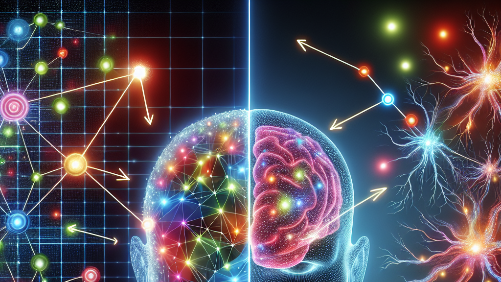
That’s where this fascinating new research comes in. Scientists have created something called a Spatial-temporal Generative Adversarial Network, or STGAN for short. Think of it as a kind of magic translator that takes a video of one kind of microscopic object and turns it into another. This process helps reveal how things inside the cell are connected, both in how they look and how they change over time.
Why does this matter? Well, by seeing more parts of the cell simultaneously, scientists could better understand how cells work, which could lead to breakthroughs in medical diagnoses and treatments. It’s like having a superpower that lets you see more than you ever could before, just by using videos instead of more colors. Imagine doctors diagnosing diseases with unprecedented precision simply by watching a video of a cell in action!
Scientists can now use video, not just colors, to see more parts of a cell at once!
FAQs
How does video-to-video translation help scientists see more cellular structures?
Video-to-video translation allows scientists to transform a video of one microscopic object into another, using a system called STGAN, which reveals more about the cell by showing spatial and temporal connections that traditional methods might miss.
What is the benefit of using STGAN in cellular research?
Using STGAN, researchers can visualize multiple cell structures simultaneously, overcoming the limitations of traditional fluorescent labeling. This provides a more comprehensive understanding of cellular interactions and dynamics.
Why are traditional fluorescent labels limited in microscopy?
Fluorescent labels are limited because each requires a distinct color that doesn’t interfere with others. As there’s only a limited number of available colors, scientists can only highlight a few structures at once, restricting the depth of cellular analysis.
Background
Microscopy traditionally uses fluorescent dyes to label different cell structures, allowing researchers to see them under a microscope. However, each type of dye emits a specific color, and the spectrum of colors is limited, which restricts the number of structures visible at one time. Generative Adversarial Networks (GANs) are powerful tools in artificial intelligence that can create new, synthetic instances of data that mimic real-world examples.
History
The exploration of cellular structures has long relied on fluorescent microscopy, a breakthrough that allowed scientists to color different parts of cells. However, the limited palette of dyes has been a constraint. The advent of artificial intelligence in image processing, particularly GANs, has opened new horizons by allowing the transformation and synthesis of visual data, leading to the development of solutions like STGAN, which can translate video data across different domains.
Based on “Revealing Microscopic Objects in Fluorescence Live Imaging by Video-to-video Translation Based on A Spatial-temporal Generative Adversarial Network” by Yang Jiao, Mei Yang, Mo Weng, available on arXiv (arxiv.org/abs/2502.16342), used under CC BY 4.0 (creativecommons.org/licenses/by/4.0/).