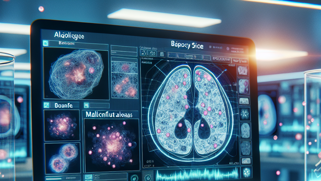



































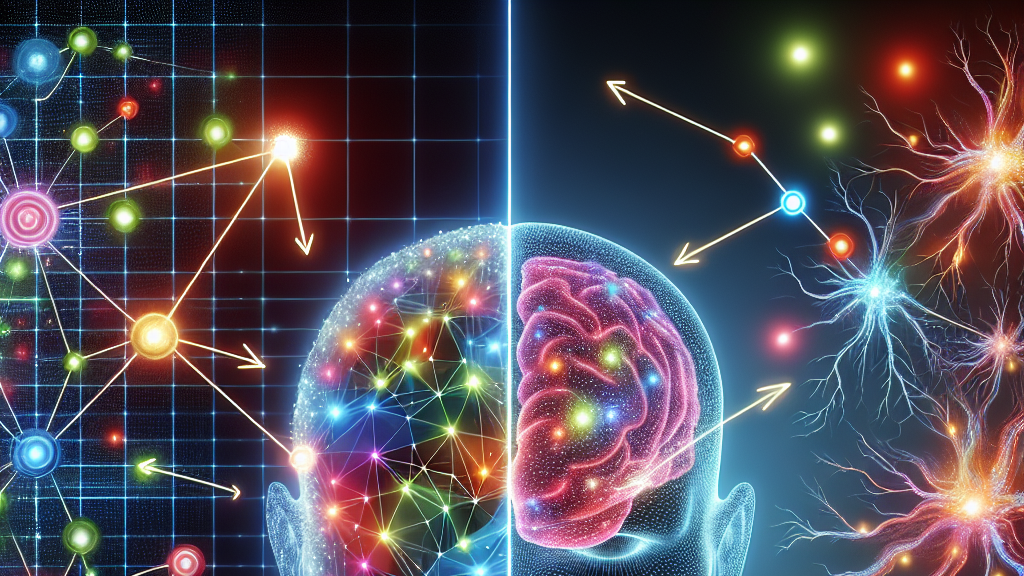








Imagine you’re at a doctor’s office, and instead of waiting weeks for your brain scan to be analyzed, a computer does it in seconds with astonishing accuracy. That’s the future vision behind new research using deep learning AI models to understand brain tissue properties from optical images, saving time and increasing reliability.
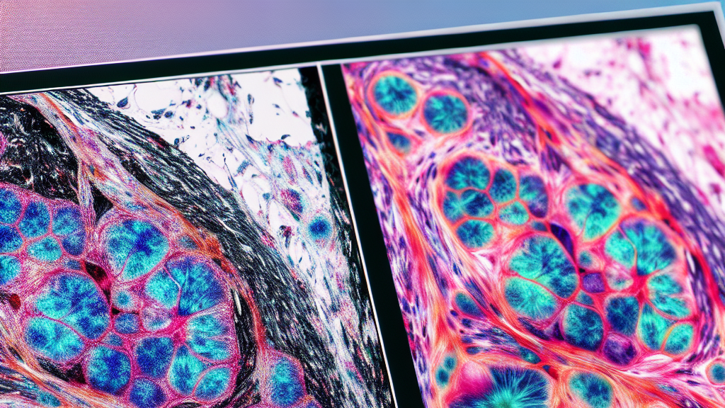
This study zeroed in on two powerful AI models, ResNet50 and DenseNet121, testing them on thousands of images from brain tissue samples. They used a clever setup called transfer learning, which means starting with a model that already knows a lot about the world, then teaching it specific tasks like recognizing patterns in brain cells. The DenseNet121 model showed it could spot these patterns with over 88% accuracy, making it a top candidate for assisting doctors.
In the future, this technology could help doctors quickly spot issues in brain scans, like tumors, leading to faster treatment plans. Imagine if every hospital used this AI; it could mean earlier diagnoses for patients and potentially save lives. This isn’t just smart science—it’s a peek into a world where machines and humans work hand-in-hand for better health outcomes.
Did you know? AI models like DenseNet121 can recognize patterns in brain tissues that are invisible to the human eye, achieving over 88% accuracy!
FAQs
How can AI improve brain scan analysis?
AI, particularly models like DenseNet121, can automatically and accurately interpret complex brain scans, saving time and reducing human error, especially in diagnosing brain diseases.
What makes DenseNet121 effective for brain tissue analysis?
DenseNet121 is highly effective because its dense connectivity allows for superior pattern recognition, even when trained on limited medical datasets, ensuring precise results.
Why use AI instead of manual analysis for brain scans?
AI can consistently interpret brain scans more quickly and with less variability between different people, making diagnoses more reliable and accessible across different healthcare settings.
Could AI technology like this be used beyond brain scans?
Definitely! AI’s ability to analyze complex patterns quickly can be adapted for various medical images, potentially transforming how we diagnose and treat diverse health issues.
What is transfer learning, and why is it used in this research?
Transfer learning involves retraining an existing AI model on new data for a specific task. This method boosts efficiency and effectiveness in learning from complex data, like brain scans, by leveraging pre-learned patterns.
Background
Optical transmission spectroscopy involves sending light through brain tissue samples to study their structural features. However, interpreting these images by eye can be tricky and varies between experts. AI, especially deep learning using convolutional neural networks (CNNs), can help—these models learn to identify intricate patterns directly from raw images, cutting down human error and time spent. The study uses two of these models, ResNet50 and DenseNet121, trained to recognize brain tissue details from a large dataset of brightfield microscopy images.
History
In the past, manual interpretation of brain scans was the norm, with experts comparing them to known samples. This process could be slow and subjective. Over the years, AI emerged as a promising tool in healthcare, offering speed and consistency. DenseNet121 and ResNet50 are advanced CNN models originally designed for general image recognition tasks; researchers now realize their potential in medical imaging, marking an evolution from basic neural networks to sophisticated, efficient AI systems for diverse applications.
Based on “Evaluation and optimization of deep learning models for enhanced detection of brain cancer using transmission optical microscopy of thin brain tissue samples” by Mohnish Sao, Mousa Alrubayan, Prabhakar Pradhan, available on arXiv (arxiv.org/abs/2505.11735), used under CC BY 4.0 (creativecommons.org/licenses/by/4.0/).