

























































Imagine going to the doctor with worries about your gut health and leaving without answers because the problem was too subtle for their tools to detect. That’s a reality many patients face, but a new technology called Multi-contrast Laser Endoscopy (MLE) could change everything. MLE is like giving doctors superhero vision to see tiny details in tissue that were previously invisible. This means potentially catching issues much earlier than before.
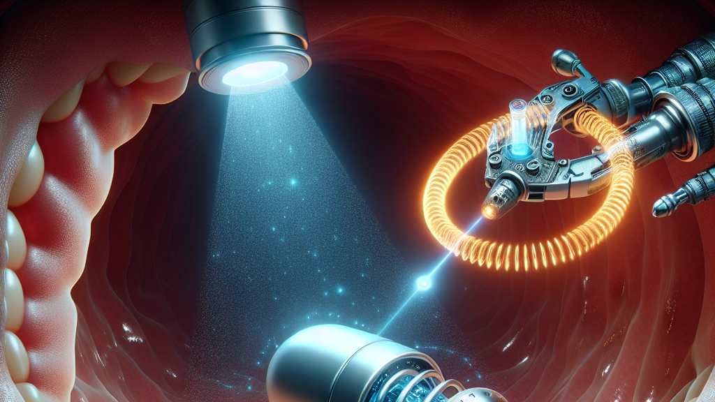
So how does this technological marvel work? MLE uses special lasers to highlight things like blood flow and tissue structure that are easily overlooked with traditional white light used in current endoscopy. It does this by rapidly changing the light’s color and direction, giving doctors a better view of what’s happening inside your digestive system. In testing, MLE has shown a dramatic improvement in contrast and color detail compared to what doctors currently use, making tricky diagnoses much easier.
Think about routine colonoscopies that might catch common polyps before they become serious health threats. With MLE, doctors could detect these much earlier and with more accuracy. Imagine a future where digestive diseases are not a constant threat looming over us, because we can catch them before they become untreatable. This isn’t just a peek into the future of medical imaging—it’s a leap forward in saving lives.
MLE technology has improved the visibility of polyps by up to three times compared to traditional imaging methods.
FAQs
What is Multi-contrast Laser Endoscopy (MLE) and how does it work?
Multi-contrast Laser Endoscopy (MLE) is a cutting-edge medical imaging technique that utilizes lasers to enhance the visibility of tissue details like blood flow and structure. It rapidly changes the light’s color and direction, providing doctors with much clearer images than traditional methods.
How does MLE improve the detection of diseases in the gastrointestinal tract?
MLE enhances the contrast and color difference of tissues, making subtle abnormalities easier to spot. In clinical tests, it has shown a three-fold improvement in contrast and a five-fold improvement in color differentiation compared to traditional methods.
What are the practical applications of MLE in everyday medical practice?
MLE could revolutionize routine procedures like colonoscopies by improving early detection of issues such as polyps, which can prevent them from developing into more serious conditions. It integrates seamlessly into existing practices, making it a practical tool for doctors.
Why is improving contrast and color detail important in endoscopy?
Better contrast and color detail allow for earlier and more accurate detection of subtle tissue abnormalities that could indicate disease. This can lead to timely treatments and better patient outcomes.
How significant is the improvement brought by MLE compared to white light imaging?
MLE provides about three times the improvement in contrast and five times the improvement in color differentiation, which is a significant leap forward in the clarity and usefulness of endoscopic images.
Background
Endoscopy is a medical procedure where doctors use a camera-equipped tool to inspect the inside of the gastrointestinal tract. Traditionally, this involves using white light to illuminate the area and identify any issues by examining tissue color, texture, and shape. However, many problems are too subtle to be detected this way. Multi-contrast Laser Endoscopy (MLE) uses lasers that can be tuned to different colors and directions to enhance these subtle features, offering a significant upgrade to traditional methods.
History
Endoscopy has been a crucial tool for gastrointestinal diagnostics for decades, primarily using white light to illuminate and examine tissues. Advances over the years have included narrow band imaging, which improves certain contrasts, but still often falls short. MLE represents a major leap, building on these advancements by using laser technology to provide a much clearer view of subtle tissue abnormalities.
Based on “Multi-contrast laser endoscopy for in vivo gastrointestinal imaging” by Taylor L. Bobrow, Mayank Golhar, Suchapa Arayakarnkul, Anthony A. Song, Saowanee Ngamruengphong, Nicholas J. Durr, available on arXiv (arxiv.org/abs/2505.10492), used under CC BY 4.0 (creativecommons.org/licenses/by/4.0/).